

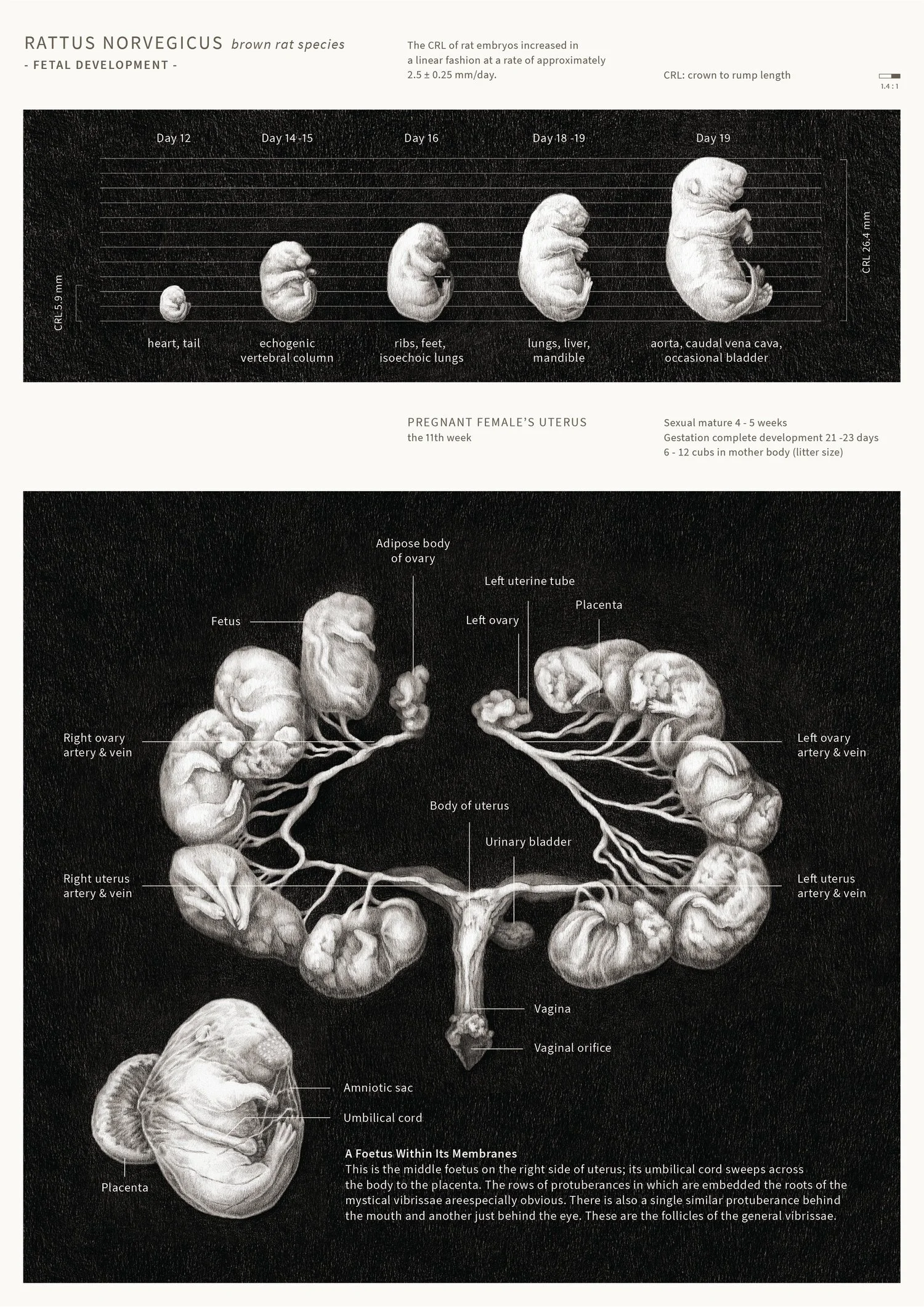
Rat fetal development
AOI - World Illustration Awards 2022:
Science & Technology Longlisted & Shortlisted
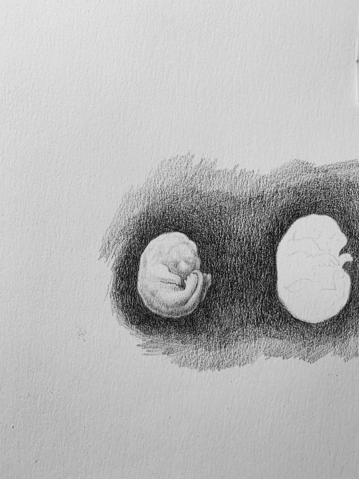
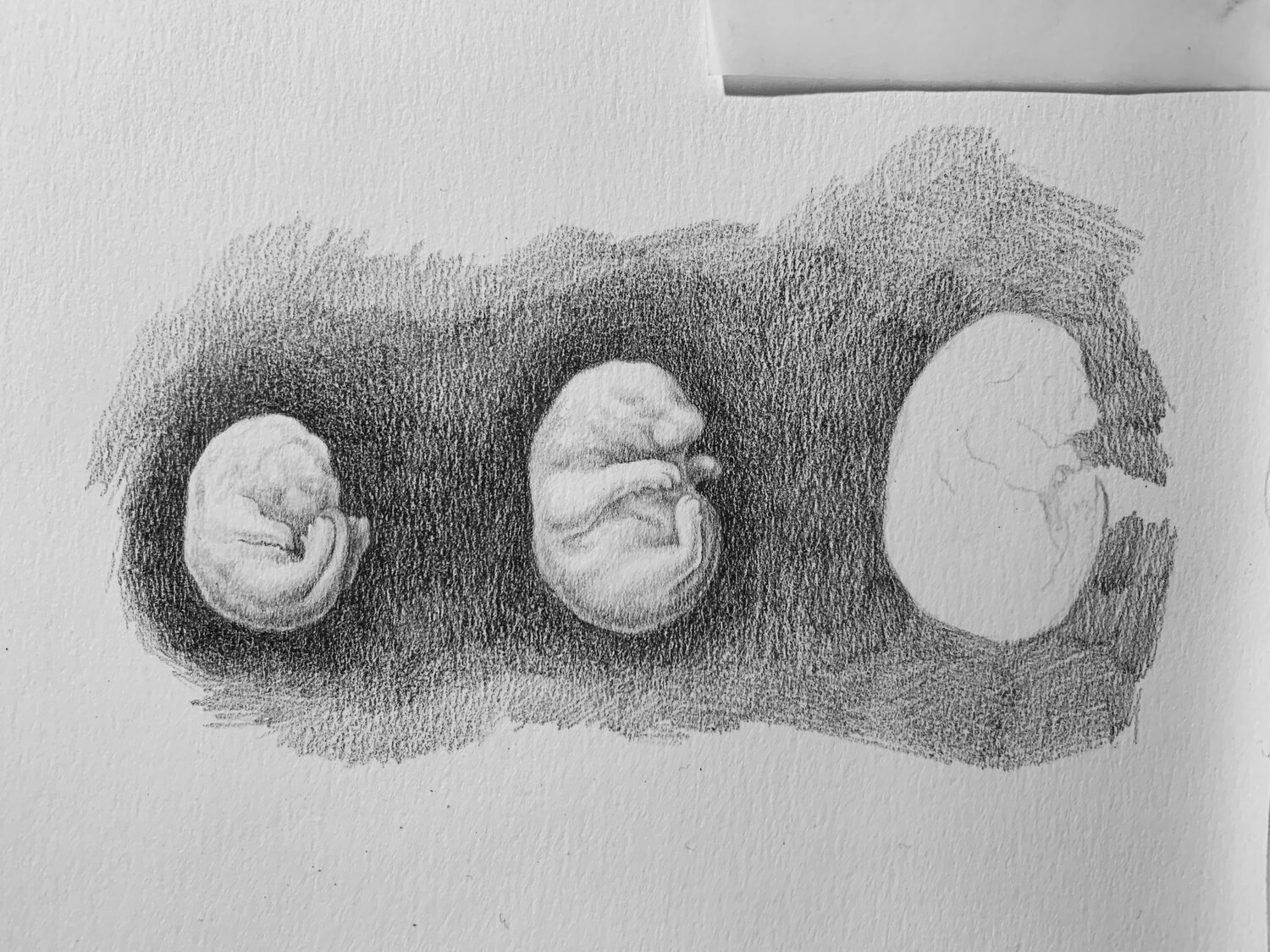
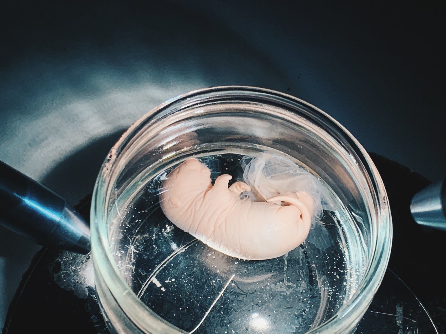
An infographic on Rattus Norvegicus’s fetal development. A visual representation on rat embryo development and its relationship with female rat’s uterus anatomy. The visual creation process contains analysing and visualising the complex data of changing CRL, organ development of rat before birth, and study anatomy through rat dissection. Information are collected through atlas, articles, and rat specimen from Maastricht University.
References:
E. M. Torres, U. M. Weyrauch, R. Sutcliffe, and S. B. Dunnett. (2008). A Rat Embryo Staging Scale for the Generation of Donor Tissue for Neural Transplantation. Department of Biosciences, Cardiff University
Kirberger RM, Bester EG, Schulman ML, Janse van Rensburg I & Hartman MJ. (2019). Ultrasonographic evaluation of fetal development in the rat. Theriogenology, 125, 24-29. PMID: 30388467
Joan R. Olds, R. J. Olds, Ronald J. Olds. A Colour Atlas of The Rat - Dissection Guide. Wolfe Medical Publications, 1979.